
EmbryLab: Build Embryo-Vision Models From Your Own Images
Label your own embryo images, accumulate a dataset as your lab works, and train a model when the data behind a label is ready.
Most AI in IVF labs today comes built into the equipment. The time-lapse system ships with its vendor’s model, trained on someone else’s images and grading conventions, and the lab uses it as delivered. It usually cannot be retrained on the lab’s own cases, and it never learns how that lab grades.
EMBRY gives clinics the other option: developing their own models, from their own images, graded by their own embryologists. The practical obstacle is the training data. A clinic-specific model needs a clinic-specific dataset — labeled, consistent, and large enough to be worth training on. Assembling one as a separate project, on top of running a lab, is not realistic for most clinics.
The dataset accumulates as the lab works
EmbryLab is built around a simple premise: the data should collect itself while the lab does what it already does.
As embryologists record development through Day 1, Day 2, Day 3 and Day 5, the images and annotations behind those entries build up per developmental day. Nothing is a separate data-entry exercise. The lab records what it records, and the dataset grows underneath.
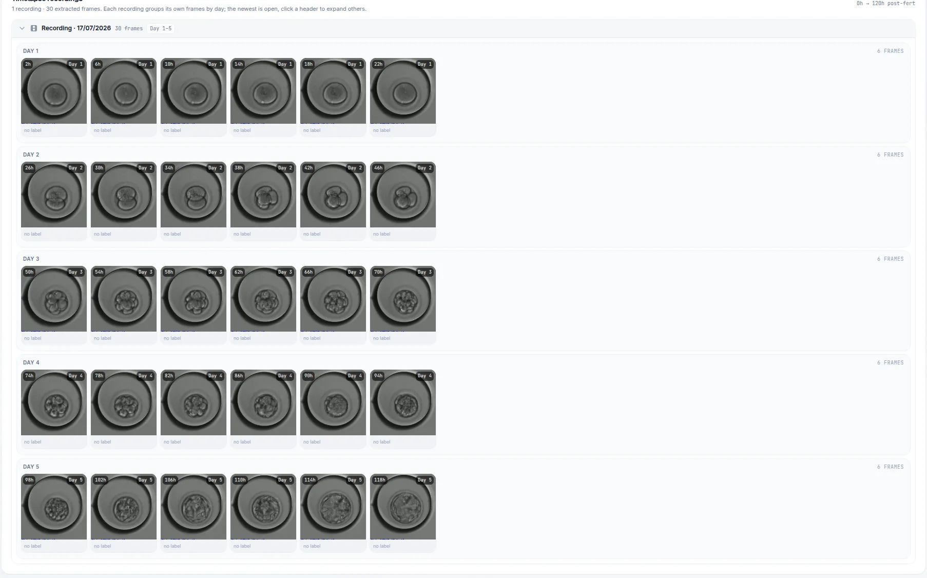
One recording, thirty frames, grouped by developmental day. Every frame is a labelling candidate — the raw material a clinic-specific model is eventually trained on.
This also means the training target is per-day rather than one general-purpose model. A Day 2 cleavage-stage image and a Day 5 blastocyst are different visual problems, and treating them as one problem is a common way to get a mediocre model.
A labeling studio inside the platform
EmbryLab includes a full annotation environment — segmentation regions, detection boxes, and keypoints — drawn directly on embryo images. Pre-installed drawing campaigns give a lab a structured starting point rather than a blank canvas and an open question about what to label.
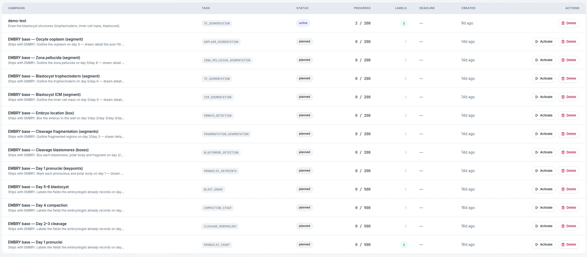
The campaigns that ship with EMBRY. Each one names a structure, a developmental day and a target — so the first question a lab faces is which to activate, not what to draw.
A trained segmentation model can then be run on an embryo’s frames, and the regions it predicts are overlaid on the image for the embryologist to check. Annotation and inference are the same loop, not two disconnected tools.
Training readiness: knowing when there is enough
The most useful thing EmbryLab does may also be the least glamorous. Each label carries a training-readiness indicator that tells you whether it has accumulated enough data to train on.
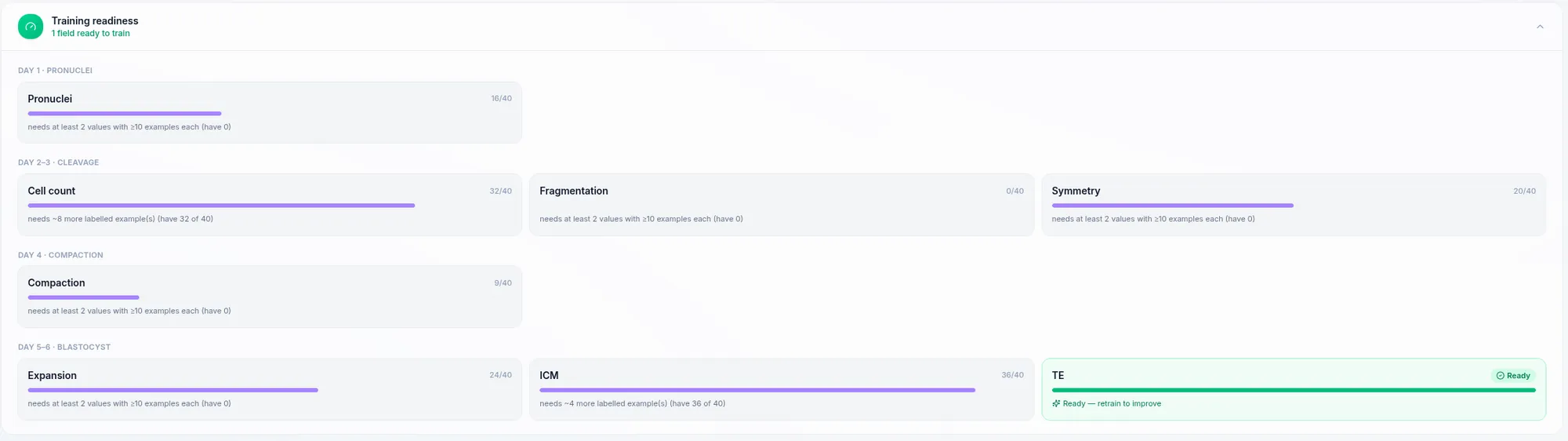
Readiness stated per field and per day. Most of these are not ready yet, and the panel says so plainly — including how many more labelled examples each one needs.
This removes a genuinely difficult judgement call. Training on a thin dataset produces a confident, useless model — and it is not obvious from the inside that this has happened. An explicit gate means a lab knows where it stands: which labels are ready, which are close, and which need more cases before anyone should train.
Not only embryo grading
The labeling and training tools are not restricted to embryo assessment. A lab can set up its own labelling campaign for a structure it wants outlined consistently — anything it can see in its own images or video — draw it, and train a segmentation model for it.
Clinics tend to have at least one of these: a recurring visual observation that matters locally, is well understood internally, and has never been captured systematically because no commercial model exists for it. This is where a lab’s actual expertise lives.
What this means for your clinic
- Your training dataset builds up through normal lab work rather than as a separate project.
- Labeling, training, and prediction happen in one loop inside the platform.
- Per-day datasets mean models suited to each developmental stage.
- A readiness gate tells you when a label can genuinely be trained, instead of leaving it to guesswork.
- You can define labelling targets specific to your own lab and train segmentation models for them.
Book a demo to see the annotation studio and readiness gate against a sample dataset.